
If membranes were able to fuse spontaneously, chaos would result. The merger of trillions of vesicles, organelles, and cells would eliminate compartmentalization, cellularity, and life itself. Fortunately, the energy barriers related to membrane deformation and fusion are high, thus two membranes cannot spontaneously merge. Membrane fusion is a key process in viral entry and reproductive biology.
Our laboratory strives to understand the molecular mechanisms that viral and cellular fusogens use to modulate multiple biological processes, such as viral-host entry and sperm-egg fusion. The overarching vision is focused on identifying and better understanding the role of membrane fusogens at the atomic level. Our primary research objectives are focused on two main areas:
a) Understand the complete molecular mechanisms and multiple functions of viral fusogens
b) Understand the diversity of cell-cell fusogens across the kingdoms of life
a) Virus entry
The entry of enveloped viruses, such as influenza A, HIV-1, SARS-CoV, and Ebola virus and others, utilize viral glycoproteins on its surface to mediate cellular attachment, tropism, and viral-host membrane fusion.
Dramatic conformational changes in the glycoproteins, triggered through pH-dependent, pH-independent, or two-step mechanisms, are necessary for fusion. We are interested in understanding the molecular mechanisms of viral-cell membrane fusion in order to develop new antivirals that block viral entry.

b) Cell-cell fusion

The fusion of membranes is also important in many biological processes, such as fertilization (sperm-egg fusion), placenta development (trophoblast fusion), skeletal muscle development (myoblast fusion), bone development (osteoclast fusion), among other processes. The mechanisms of cell-cell fusion are poorly defined and have generally been based on the mechanisms employed by viruses for fusion. Recently, it seems some cell-cell fusion processes may be evolutionarily related to the viral glycoproteins (for example C. elegans EFF-1 protein, and syncytin-1/syncytin-2 in placenta development). We are interested in understanding the various mechanism of cell-cell fusion and how it relates to virus-cell fusion.
Our approach
Using our structural models as a molecular blueprint, we aim to develop new therapeutic strategies.
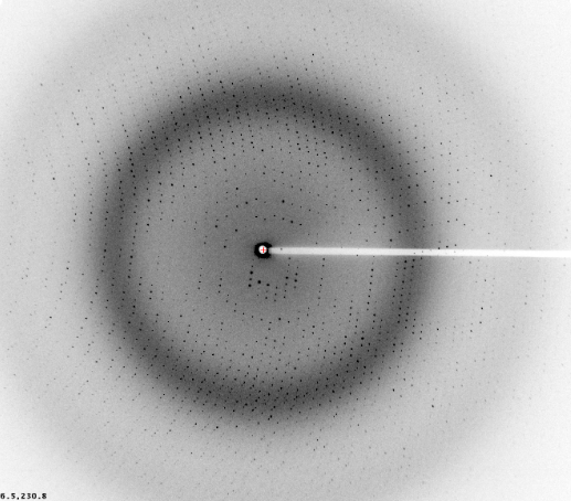
The main tool we employ to understand cellular and viral protein function is X-ray crystallography and cryo-EM. Crystallographic analysis of viral and cellular surface glycoproteins has offered a tremendous wealth of insights into recognition, entry, fusion, restriction, and pathogenesis. We also combine X-ray crystallography and cryo-EM with other biophysical and biochemical techniques such as small angle X-ray scattering (SAXS) and deuterium exchange mass spectrometry (DXMS) to reach the higher hanging fruits. Once structures are determined, questions and hypotheses arising will be subsequently tested using biochemical, immunological and virological techniques. Our long-term goals are to understand the fundamental principles behind key biological processes, identify new targets and provide a template for the design and development of new therapeutic strategies.